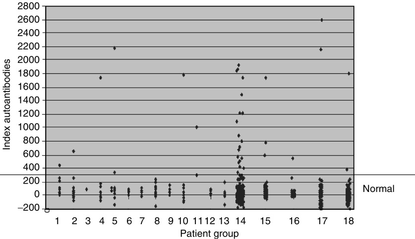
Figure 4.
PDXP autoantibody analysis using immunoprecipitation assays. The figure shows the distribution of index values for the different groups of subjects used in this study. PDXP antibody reactivity is given as indices relative to a positive (patient serum) and negative control (pool of sera from healthy individuals) ((cpm subject X−cpm negative standard)/(cpm positive standard−cpm negative standard) × 1000). The cutoff value for identifying positive samples for autoantibodies against PDXP was 279. Lanes 1–14: sera from patients with tumours of different origins. 1: lymphoma, 2: mammary, 3: bladder, 4: ovary, 5: uterus, 6: testis, 7: skin (malignant melanoma), 8: prostate, 9: kidney, 10: colon, 11: bile duct, 12: rectum, 13: other, 14: lung. Lanes 15–18: sera from patients with other autoimmune diseases and controls. 15: Addison's disease or APS type II, 16: type 1 diabetes, 17: multiple sclerosis and 18: blood donors.

