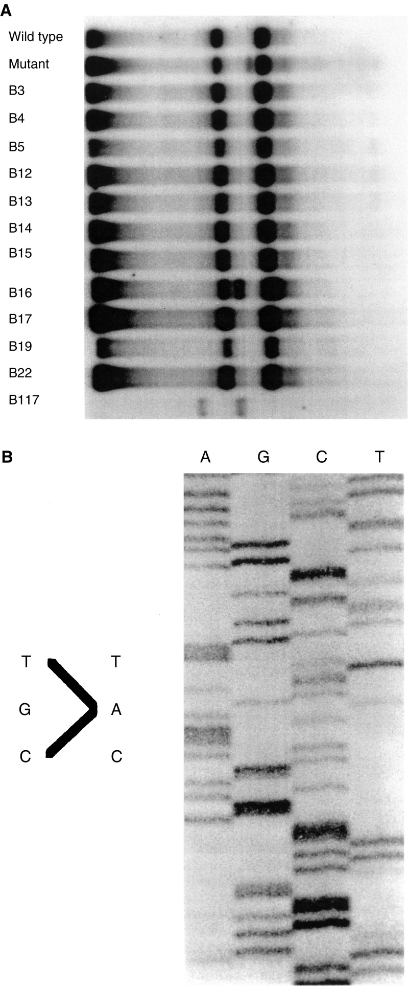
Figure 3.
(A) A SSCP analysis of exon 8 of the p53 gene run on 12% polyacrylamide gel with 1% glycerol at 10 W for 16 h. Note band shift in lane marked B 16 (indicating a p53 alteration). The shift is a result of conformational changes induced by the point mutation identified in the sequencing gel in (B). (B) DNA sequence analysis of exon 8 for sample B 16 indicating base pair substitution at codon 273 changing an arginine residue to histidine. Nucleotide base pairs: A=adenine; G=guanine; C=cytosine; T=thymine.

