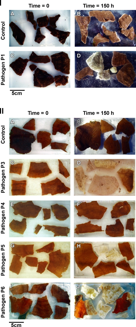
Figure 3. Inoculation experiment II:
I A–B. Montipora aequituberculata coral fragments in un-inoculated control treatment (t = 0h and t = 150h). I C–D. M. aequituberculata coral fragments exposed to 1×10⁶ cells ml −1 of culture P1 (t = 0h and t = 150h). II A–B. Pachyseris speciosa coral fragments in un-inoculated control treatment (t = 0h and t = 150h). II C–D. P. speciosa coral fragments exposed to 1×10⁶ cells ml−1 of culture P3 (t = 0h and t = 150h). II E–F. P. speciosa coral fragments exposed to 1×10⁶ cells ml−1 of culture P4 (t = 0h and t = 150h). II G–H. P. speciosa coral fragments exposed to 1×10⁶ cells ml−1 of culture P5 (t = 0h and t = 150h). II I–J. P. speciosa coral fragments exposed to 1×10⁶ cells ml−1 of culture P6 (t = 0h and t = 150h).

