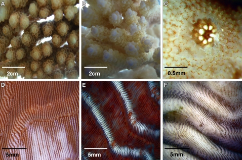
Figure 5. Disease progression:
A. Acropora hyacinthus fragmennt inoculated with 1×10⁶ cells ml−1 of culture P2 (t = 0h). B. Loss of Symbiodinium from A. hyacinthus inoculated with 1×10⁶ cells ml−1 of culture P2 (t = 12h). C. Polyp and surrounding tissue-loss of Symbiodinium from A. hyacinthus inoculated with 1×10⁶ cells ml−1 of culture P2 (t = 12h). D. Loss of Symbiodinium cells from Pachyseris speciosa inoculated with 1×10⁶ cells ml−1 of culture P3 (t = 12h). E. Tissue lesions on P. speciosa inoculated with 1×10⁶ cells ml−1 of culture P3 (t = 24h). F. Exposed skeleton on P. speciosa inoculated with 1×10⁶ cells ml−1 of culture P3 (t = 60h).

