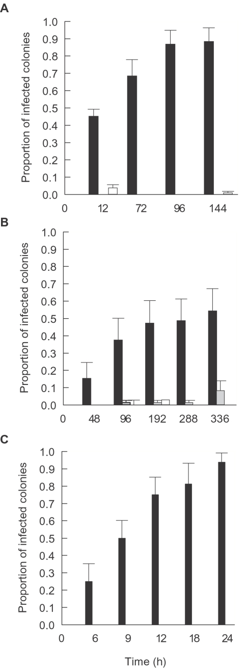
Figure 6. Disease transmission:
A. vMean proportion of infected Pachyseris speciosa coral fragments displaying WS signs following exposure to cultures of P3–P6 in comparison to proportions in inoculated and un-inoculated control treatments. B. Mean proportion of infected Montipora aequituberculata coral fragments displaying WS signs following exposure to culture of P1 in comparison to proportions in inoculated and un-inoculated control treatments. C. Mean proportion of infected Acropora cytherea coral fragments displaying WS signs following exposure to culture P2 in comparison to proportions in inoculated and un-inoculated control treatments. ▪-Coral fragments inoculated with 1×10⁶ cells ml−1 of putative pathogen cultures.  -Coral fragments inoculated with 1×10⁶ cells ml−1 culture of non-pathogen isolates. □-Coral fragments without inoculation. Time represents hours (h) following exposure. Bars = Standard errors.

