Fig. 4.
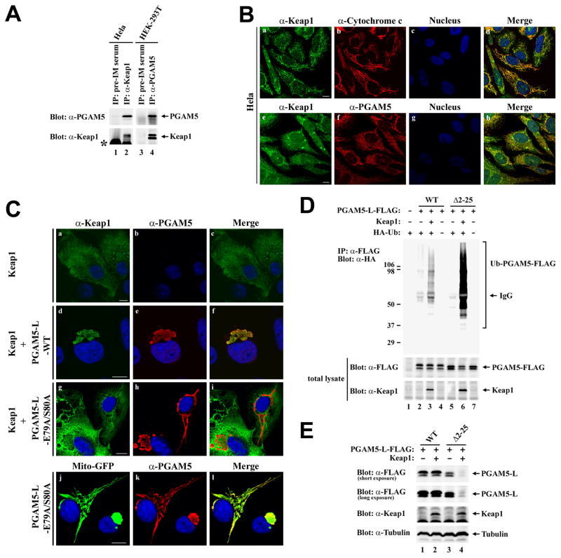
PGAM5 recruits Keap1 to mitochondria. (A) Anti-Keap1 immunoprecipitates from 6 mg of Hela cell lysates (lane 2) and anti-PGAM5 immunoprecipitates from 8 mg HEK-293T cell lysates (lane 4) were subjected to immunoblot analysis with affinity-purified anti-PGAM5 (top panel) and anti-Keap1 antibodies (bottom panel). Pre-immune rabbit and chicken serum were used as negative controls for the anti-Keap1 and anti-PGAM5 antibodies, respectively (lanes 1 and 3). The asterik indicates the presence of rabbit IgG in lanes 1 and 2. (B) Hela cells were grown on coverslips in 24-well plates. The cellular localization of the endogenous Keap1 protein was determined by indirect immunofluorescence with affinity-purified anti-Keap1 antibodies against the full-length Keap1 (panels a and e). Mitochondria were visualized by indirect immunofluorescence using anti-Cytochrome c antibodies (panel b). PGAM5 was detected by indirect immunofluorescence using affinity-purified anti-PGAM5 antibodies (panel f). Nuclei were stained with Hoechst 33258. Merged images were shown on the right (panels d and h). (C) COS1 cells were transfected with expression vectors for Keap1, untagged wild-type PGAM5-L, mutant PGAM5-L-E79A/S80A and Mito-GFP as indicated. The cellular localization of the ectopic Keap1 and PGAM5-L proteins was determined by indirect immunofluorescence with anti-Keap1 (panels a, d, and g) and anti-PGAM5 antibodies (panels b, e, h and k). Mito-GFP was visualized by direct fluorescence (panel j). Nuclei were stained with Hoechst 33258. Merged images are shown on the right (panels c, f, i and l). (D) COS1 cells were transfected with expression vectors for HA-Ub (0.4 μg), Keap1 (0.4 μg), and C-terminal FLAG-tagged wild-type or mutant PGAM5-L proteins (0.2 μg) as indicated. Anti-FLAG immunoprecipitates (IP) were analyzed by immunoblot with anti-HA antibodies (top panel). Total lysates were analyzed by immunoblot with anti-FLAG and anti-Keap1 antibodies (bottom two panels). (E) Hela cells were transfected with expression vectors for Keap1 (0.5 μg), and C-terminal FLAG-tagged wild-type or mutant PGAM5-L proteins (0.5 μg) as indicated. Total lysates were analyzed by immunoblot with anti-FLAG, anti-Keap1, and anti-Tubulin antibodies. White bars = 10 microns.

