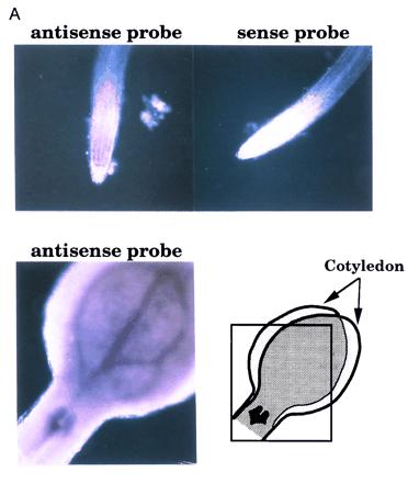
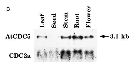
Figure 5.


The expression pattern of AtCDC5. (A) In situ analysis of AtCDC5. Using antisense and sense RNA probes, whole mount in situ hybridization was performed. Microscopic views of root tips and a vegetative shoot apex are shown. Note that high background signals in cotyledons was due to their overlapping as indicated. (B) Northern blot analysis of AtCDC5 and CDC2a. RNAs from leaves, developing siliques, shoots, roots, and flowers were hybridized with the AtCDC5 cDNA probe or the CDC2a cDNA probe (3).
