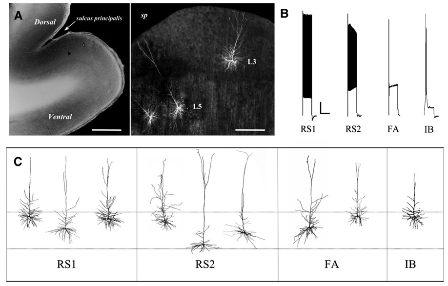
FIG. 1.
A, left: photomicrograph of a typical prefrontal cortex (PFC) slice, Scale bar: 1.2 mm; right panel: higher-magnification view of a PFC with 3 representative biocytin-filled pyramidal cells (2 in layer 5, 1 in layer 2/3). Scale bar: 300 µm. B: distinctive action potential (AP) firing patterns elicited by a 2-s, 280-pA depolarizing current step in the 4 types of layer 5 pyramidal cells. Note the progressive decrease in AP amplitude and increase in AP threshold in regular-spiking slowly adapting type-2 (RS2) and regular-spiking fast-adapting (FA) but not regular-spiking slowly adapting type-1 (RS1) cells, and the rapid rate of adaptation in FA cells only. C: diagram of representative reconstructed layer 5 pyramidal cells of each electrophysiological class. - - -, approximate boundaries of layer 5 (~900–1,500 µm deep to the pial surface).

