
Figure 3.
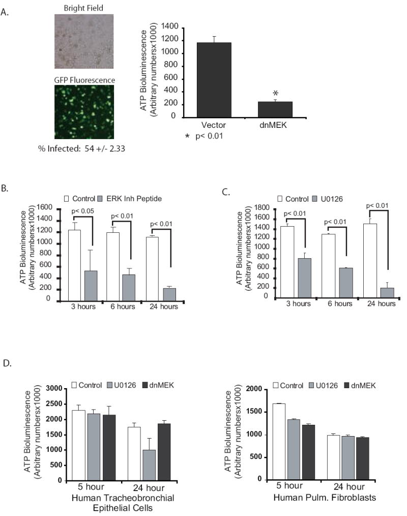
ERK inhibition decreases ATP specifically in alveolar macrophages. 3A. Human alveolar macrophages were cultured (1×10⁶/ml in 96 well tissue culture plates). At time 0 the cells were infected with Ad dnMEK as described in the methods. Control cells were infected with an adenovirus vector expressing GFP. At 6 hours post infection, cells were lysed and total ATP levels analyzed as described. The data represents three separate experiments. Significance was determined using a nonpaired t test. 3B. Human alveolar macrophages were cultured (1×10⁶/ml in 96 well tissue culture plates) with and without a peptide ERK inhibitor (50 uM). At 3, 6 and 24 hours post inhibition, cells were lysed and total ATP levels analyzed as described. The data represents three separate experiments. Significance was determined using nonpaired t tests. 3C. Human alveolar macrophages were cultured (1×10⁶/ml in 96 well tissue culture plates) with and without the MEK inhibitor, U0126 (20 uM). At 3, 6 and 24 hours post inhibition, cells were lysed and total ATP levels analyzed as described. The data represents three separate experiments. Significance was determined using nonpaired t tests. 3D. Primary human tracheobronchial cells and primary human fibroblasts were grown in 96 well plates and then exposed to the ERK/MEK inhibitor, U0126 (20 uM) for various times. ATP was measured as described and is presented as arbitrary bioluminescence numbers. The data represents three experiments.