
Figure 5.
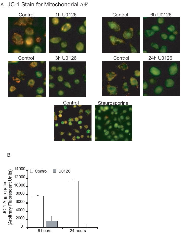
ERK inhibition causes loss of mitochondrial membrane potential (mitΔψ) in human alveolar macrophages. 5A. Human alveolar macrophages were cultured (1×10⁶/ml in 6 well tissue culture plates or in 2 chamber microscope slides) with and without U0126 (20 uM) or staurosporine (1 uM). At the end of the incubation period, the mitochondrial stain JC-1 was added as described in the methods. Cultures were incubated a further 30 minutes and then examined with fluorescence microscopy. Photomicrographs were obtained. Red/orange stain denotes intact mitochondria with no disruption of membrane potential. Green staining denotes loss of mitΔψ. 5B. Identical experiments were performed in 96 well tissue culture plates and red/orange stain quantified using a fluorescence plate reader. The graph represents arbitrary units at an excitation of 535 nM and an emission of 590 nM.