
Figure 7.
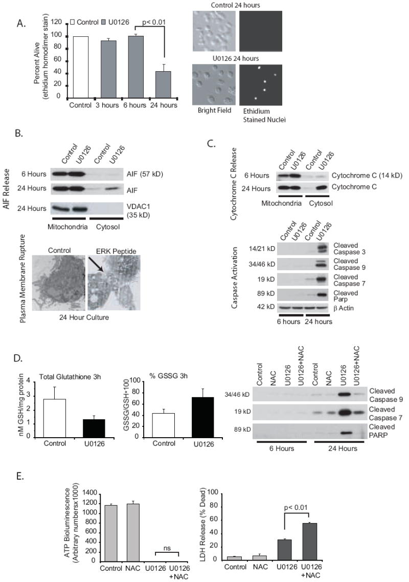
ERK inhibition induces both apoptotic and necrotic pathways. 7A. ERK inhibition leads to alveolar macrophage death. Human alveolar macrophages were cultured (1×10⁶/ml in 6 well tissue culture plates) with or without U0126 (20 uM) for 3, 6 or 24 hours. At the end of each time point, EthD-1 entry was evaluated. Dead cells were counted by examining each cell for red nuclear staining. A total of 300 cells were examined for each data point. The data is graphed as percentage of cells that exclude EthD-1. The graph represents data from three separate experiments. Significance was determined using a nonpaired t test. 7B. ERK inhibition results activation of necrotic pathways. Alveolar macrophages were cultured for 6 or 24 hours (1×10⁶/ml in 6 well tissue culture plates) with or without U0126 (MEK inhibitor, 20 uM). The blots show mitochondrial and cytosolic protein fractions analyzed (Western analysis) for AIF. The 24 hour blot was stained with VDAC as a control for the mitochondrial isolation. Also shown is TEM demonstrating loss of plasma membrane integrity after 24 hours with an ERK inhibitory peptide (50 uM). Cells were fixed overnight with 2.5% glutaraldehyde in 0.1 M cacodylate buffer. They were then processed for TEM as described in the Methods section. 7C. ERK inhibition activates apoptotic pathways. Alveolar macrophages were cultured for 6 or 24 hours (1×10⁶/ml in 6 well tissue culture plates) with or without U0126 (MEK inhibitor, 20 uM). The first blot shows mitochondrial and cytosolic protein fractions analyzed (Western analysis) for cytochrome C. The other blots show total cell lysates stained for cleaved caspase 3, 7 and 9 and cleaved PARP. Equal loading was determined by staining identical blots for β actin. 7D. ERK inhibition increases ROS in alveolar macrophages. Alveolar macrophages were cultured for 3 hours (1×10⁶/ml in 6 well tissue culture plates) with or without U0126 (MEK inhibitor, 20 uM). Frozen cell pellets were used to measure GSH (non-oxidized glutathione) and GSSG (oxidized glutathione) levels. Data is expressed as either total GSH or percentage GSSG (GSSG/GSH × 100). In addition the effect of blocking oxidant increases with NAC (1 mM) on caspase activity was evaluated by Western analysis of whole cell lysates at 6 and 24 hours. 7E. Inhibition of caspase activity does not prevent ERK inhibition-induced loss of ATP and decreased survival. Human alveolar macrophages were cultured (1×10⁵/100 ul in 96 well tissue culture plates) with or without U0126 (20 uM) for 24 hours. At the end of the incubation time ATP levels were measured as described in the methods and LDH release (as described in the methods) was measured as a marker of plasma membrane permeability. NAC did not prevent either the loss of ATP or lass of plasma membrane integrity by ERK inhibition.