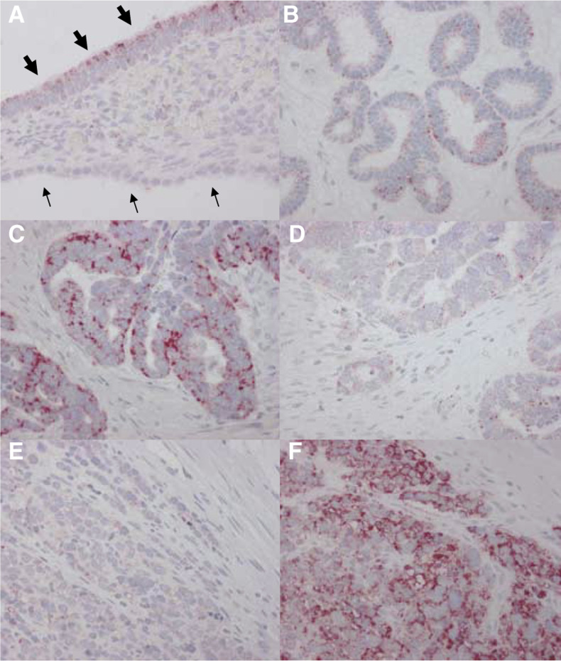
Figure 1.
Expression of PLK1 and PLK3 in ovarian tissue specimen. (A) Normal ovarian surface epithelium without significant PLK1 positivity (small arrows) and moderate PLK1 positivity in an adjacent serous cystadenoma (bold arrows). (B) Serous borderline tumour of the ovary showing only weak scattered expression of PLK3. This tumour was scored as PLK3 negative. (C, D) Serous ovarian carcinoma with strong expression of PLK1 in more than 80% of tumour cells (C), while the same tumour showed only low expression for PLK3 (D). (E, F) Undifferentiated ovarian carcinoma staining strongly positive for PLK3 (F) but not for PLK1 (E).

