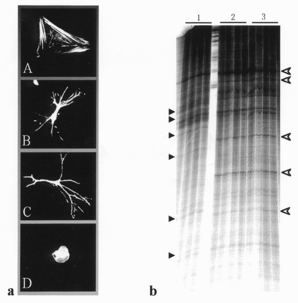
Figure 1.

Alteration in cell shape (a) and gene expression (b) by ECM in cultured HSCs. Fig. 1a shows HSC shape after culturing on polystyrene surface (A), on type I collagen gel (B), in type I collagen gel (C), or on Matrigel (D). Fig. 1b indicates a representative PCR-DD showing differentially expressed transcripts in HSCs cultured on polystyrene (1), on type I collagen gel (2), or in type I collagen gel (3). Black and open arrowheads indicate the PCR products derived from down- and up-regulated transcripts, respectively, by extracellular type I collagen.
