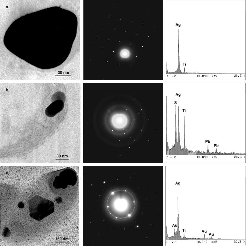
Figure 3.
Crystal structure analysis. (a) Regularly shaped nanocrystalline Ag particle taken from a thin, unstained section of a P. stutzeri AG259 cell, with a corresponding EDX spectrum (Right) and its electron diffraction pattern (Center) indicating elemental crystalline silver. (b) Second crystal type embedded in the periplasmic space of the cell. EDX spectrum and electron diffraction indicate monoclinic Ag2S. (c) A third type of crystal taken from a whole cell. The crystal structure is not yet clear. The electron diffraction pattern is not consistent with the pattern of elemental silver, whereas the EDX spectrum shows only Ag in considerable amounts.

