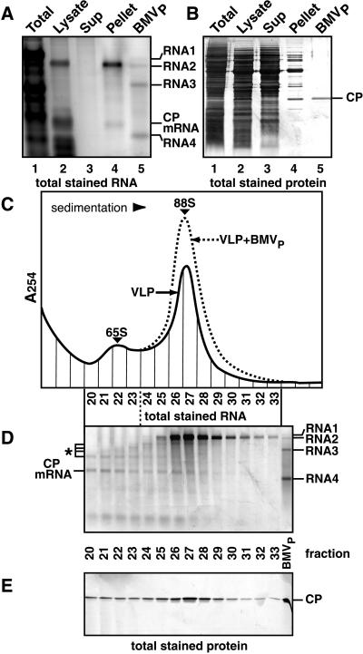
Figure 2.
Analysis of VLPs from yeast expressing BMV RNA2 and CP. (A) RNA analysis. RNA from the indicated fractions (see text) was electrophoresed through 1% agarose, stained for total RNA, and imaged by laser fluorometry. BMVP RNA (lane 5) was included as a control. The indicated bands in lanes 2 and 4 were identified as CP mRNA and RNA2 by hybridization to CP and RNA2 probes. (B) Protein analysis. Total protein was electrophoresed and visualized by silver staining. BMVP CP (rightmost lane) was included as a control. (C) Sucrose gradient sedimentation. The resuspended pellet in lanes 4 of A and B was centrifuged through a 5–30% linear sucrose gradient, and gradient fractions were analyzed by UV absorption at 254 nm. Sedimentation profiles of yeast-derived VLPs alone (solid curve) and with BMVP added (dotted curve) are shown. (D) RNA analysis of sucrose gradient fractions, as in A. BMVP RNA (right lane) was used as a control. RNA2, RNA2 fragments (*), and CP mRNA were identified by hybridization to CP- and RNA2-specific probes. (E) Protein analysis of sucrose gradient fractions, as in B.

