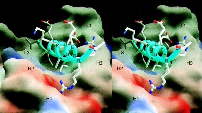
Figure 2.
Molecular surface representation of the scFv C219 (molecule II) binding site with the bound α-helical P-glycoprotein epitope peptide. The molecular surface is colored for electrostatic potential (red for negative charge, blue for positive charge). Peptide residues and the approximate locations of C219 heavy (H) and light chain (L) hypervariable loops are indicated. Fig. 2 was produced with the program grasp (32).

