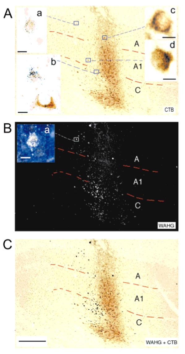
Fig. 4.

Sagittal section through the lateral geniculate nucleus of a postnatal day 16 cat (K137) viewed in brightfield for cholera toxin B (CTB) labeling (A) and in darkfield for wheat germ apo-horseradish peroxidase gold (WAHG) labeling (B). CTB was injected into ipsilateral eye patches in cortex, and WAHG was injected into contralateral patches. C: A superimposition in Photoshop of the brightfield image and the inverted darkfield image. The insets show, at high magnification, the appearance of neurons retrogradely labeled with CTB (inset c), with WAHG (insets a,b in A, a in B; insets a show the same cell in brightfield and darkfield, respectively). Double-labeled neurons are shown at high magnification in A, inset b (bottom neuron), and inset d. Scale bars = 500 μm for A-C; 10 μm for insets a-d.
