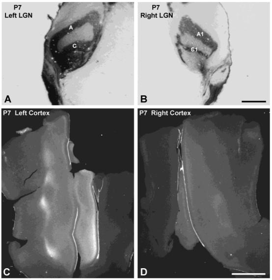
Fig. 7.

Postnatal day 7 (P7) cat. A,B: Autoradiographs of coronal lateral geniculate nucleus (LGN) sections viewed with brightfield microscopy, showing dark label concentrated in appropriate laminae after [3H]proline injection into the right eye. C,D: Autoradiographs of single, flatmounted sections of visual cortex, viewed with darkfield microscopy, showing label in layer IV. The label, which appears bright in darkfield, was stronger in the left cortex. There were no columns apparent in either hemisphere, although the LGN laminae were distinct at this age. Scale bars = 1 mm in A,B; 5 mm in C,D.
