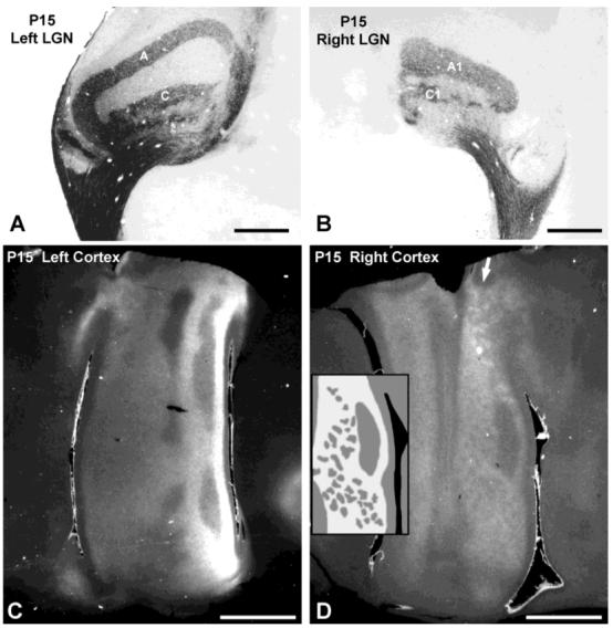
Fig. 8.

Postnatal day 15 (P15) cat. A,B: Autoradiographs showing mature pattern of lamina specific label in lateral geniculate nucleus (LGN). C,D: In the contralateral (left) cortex, the ocular dominance columns are visible but difficult to illustrate. In the ipsilateral (right) cortex, they are much clearer. Areas of bright label in (D) represent zones receiving input from the ipsilateral eye; regions of darker label receive input predominately from the contralateral eye. Arrow indicates coarser columns in area 18. Inset in D is a drawing of the ocular dominance columns in the portion of area 17 immediately to the right; scale is the same as the photograph. Scale bars = 1 mm in A,B; 5 mm in C,D.
