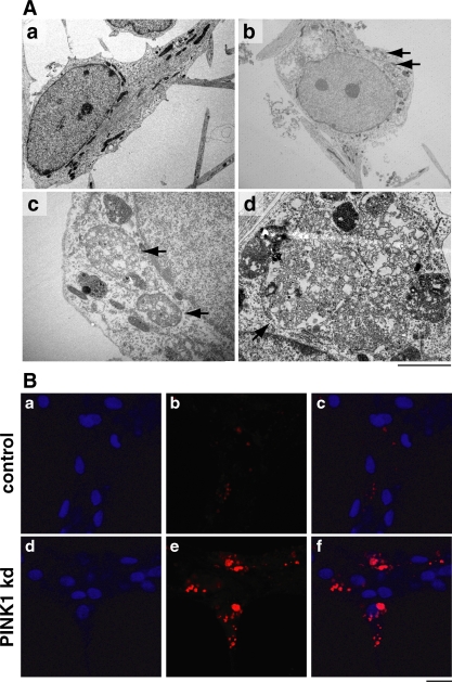
Figure 7. Abnormal lysosomal morphology in PINK1 kd neurons.
A) TEM images of vesicular aggregates within surviving aged human neurons lacking PINK1 at dd47. A control neuron is shown in panel A. Panels b-d show aggregates in PINK1 kd neurons, panel c is an aggregate enlarged from b. Scale bars; a, b; 20 µM, d; 2 µM. B) Immunofluorescence images of aged (dd47) control human neurons (panels a-c) and PINK1 kd neurons (d–f) showing lysosomal morphology (red staining; panels b and e). Nuclei are shown in blue (panels a and d), merged images presented in panels c and f. Large and multiple aggregates seen only in PINK1 kd neurons are positive for the dye Lysotracker (panel d). ,Scale bar = 20 µM

