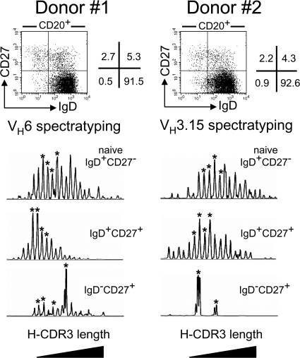
Figure 1.
H-CDR3 spectratypes of the VH6 or VH3.15 transcripts expressed by the blood B cell subsets of two 11-mo-old children. Naive IgD+CD27−, IgD+CD27+, and switched IgD−CD27+ B cells were sorted from the blood samples of donors 1 and 2. Total RNA from each cell fraction was reverse transcribed and VH6 or VH3.15 μ or γ transcripts were amplified by PCR, using a seminested strategy (see Materials and methods). The PCR products were labeled by a run-off reaction with specific fluorescent VH-FR3 primers, and subjected to electrophoresis on an automated sequencer. The resulting size distribution of the peaks directly reflects the size distribution of H-CDR3 for the given transcripts. Peaks identified by an asterisk were further sequenced to evaluate intrapeak clonal diversity.

