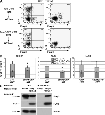
Figure 5.
Regulation of RORγt+ T cells by Foxp3. (A and B) Bone marrow cells isolated from CD45.2+Rorc(γt)-GfpTG (WT) or Foxp3sf × Rorc(γt)-GfpTG (Scurfy) mice were mixed with bone marrow cells isolated from CD45.1+ C57BL/6 mice and injected into irradiated lymphopenic mice. 6 wk after transfer, cells were isolated from the spleen, LN, and lung and restimulated in vitro with PMA/ionomycin for 5 h and subjected to intracellular staining for GFP, IL-17, Foxp3, and IL-10. (A) Spleen cells are gated on GFP+TCR-β+ cells. Numbers indicate percent cells in quadrants. Reconstitutions with CD45.1+ and CD45.2+ cells were comparable in all chimeras. Results are representative of two independent experiments. (B) Histograms report percent GFP+TCR-β+ cell subsets in total CD45.2+ T cells (see Fig. 4 C). *, P < 0.05 as compared with WT. (C) Immunoprecipitation of RORγt and Foxp3. Foxp3 was expressed in HEK293T cells alone or together with N-terminally FLAG-tagged RORγt. Total cell lysates or immunoprecipitates (IP) using anti-FLAG antibody were blotted and probed with antibodies to Foxp3, FLAG, or β-actin. Results are representative of two independent experiments.

