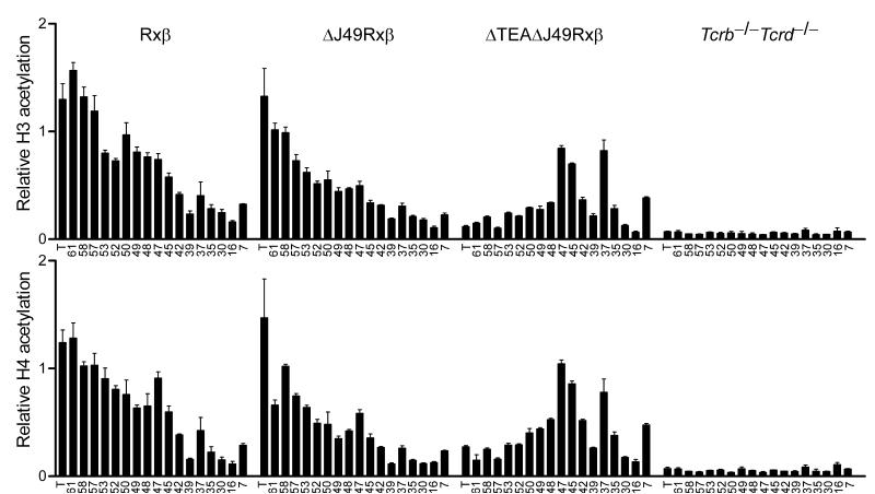
Figure 6.
Jα array histone modifications in TEA and Jα49 promoter-deleted mice. Histone H3 and H4 acetylation was analyzed by chromatin immunoprecipitation from mononucleosomes prepared from thymocytes of Rxβ, ΔJ49Rxβ, and ΔTEAΔJ49Rxβ mice, and as a control, from splenocytes of Tcrb-/-Tcrd-/- mice. Sites analyzed were situated in the TEA exon (T) or at individual Jα segments (numbered). Bound and input fractions were quantified using real time PCR and ratios of bound/input were normalized to that for B2m in each sample. The data represent the mean ± SEM of triplicate PCR reactions.

