
Fig. 2. Distribution of AR-ir axons in the rat forebrain and brainstem.
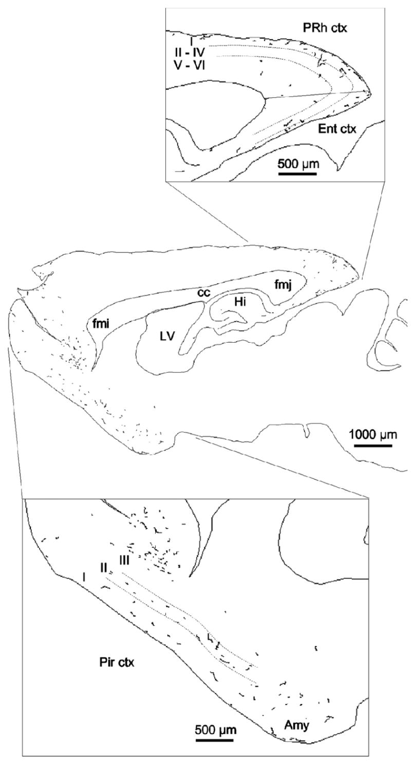
This map was produced using NeuroLucida software (MicroBrightField Inc.) and illustrates a single 40-μm parasagittal section, 2.9 mm lateral to bregma. The entire section was scanned at x40, and all AR immunoreactive axons observed were traced. An adjacent Nissl-stained section was used to delineate cortical layers. Amy, Amygdala; cc, corpus callosum; ctx, cortex; Ent, entorhinal cortex; fmi, forceps minor of the corpus callosum; fmj, forceps major of the corpus callosum. Hi, hippocampus; LV, lateral ventricle; Pir, piriform cortex; PRh, perirhinal cortex. Reproduced from (DonCarlos, et al, 2003), Copyright 2003, The Endocrine Society.