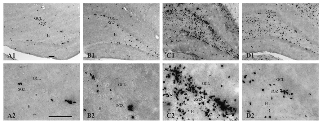
Figure 3.
BrdU-immunopositive cells (i.e., newly generated cells) in the dentate gyrus and hilus 16 days following saline treatment (A, CON; B, SUP) or KA-induced SE (C, CON; D, SUP). Note that the number of BrdU-labeled cells significantly increased 16 days after SE for both diet groups, but that this seizure-induced proliferative response was markedly attenuated in SUP rats (D) in comparison to CON rats (C). Photomicrographs in the top row were taken with a 10x objective and the bottom row was taken with a 40x objective. Bars indicate 50 µm. GCL, granule cell layer. SGZ, subgranular zone. H, hilus.

