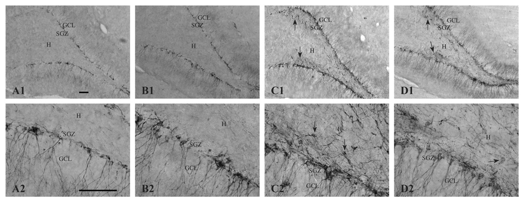
Figure 5.
DCX-immunopositive neurons (i.e., newly generated neurons) in the dentate gyrus and hilus 16 days following saline treatment (A, CON; B, SUP) or KA-induced SE (C, CON; D, SUP). The number of DCX-labeled cells significantly increased 16 days after SE for both CON (A, C) and SUP (B, D) rats. Note that in KA-treated rats of both diet groups (C, D), DCX-positive neurons were aberrantly located in the hilus and exhibited abnormal morphological features, such as horizontally oriented cell bodies and processes (indicated by arrows). Photomicrographs in the top row were taken with a 10x objective and the bottom row was taken with a 40x objective. Bars indicate 50 µm. GCL, granule cell layer. SGZ, subgranular zone. H, hilus.

