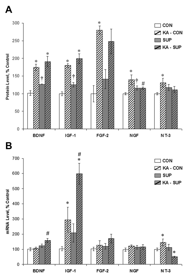
Figure 8.
Comparison between CON (white bars) and SUP (grey bars) rats in growth factor protein (A) and mRNA (B) levels (mean ± SEM percent of control levels) in the intact hippocampus (open bars) and 16 days following KA-induced SE (hatched bars). Protein levels were quantified using ELISA. SE led to a significant increase in BDNF, IGF-1, FGF-2, NGF, and NT-3 protein levels in CON rats, but a significant increase in only BDNF and IGF-1 protein levels was noted for SUP rats. Baseline protein levels of BDNF, IGF-1, and NGF were significantly higher in SUP rats. SUP rats also exhibited significant increases in BDNF and IGF-1 mRNA and a decrease in NT-3 mRNA 16 d following SE, whereas CON rats showed SE-induced increases in IGF-1 and NT-3 mRNA. * statistically different from within-diet saline-treated group; # statistically different from KA-treated CON; † statistically different from saline-treated CON.

