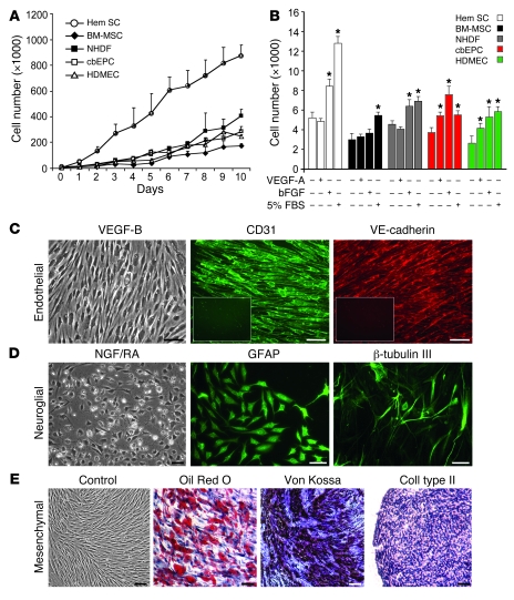
Figure 2. In vitro growth and multilineage differentiation of HemSCs.
(A) Proliferation of HemSCs in EBM-2/20% FBS over 10 days compared with normal endothelial and mesenchymal cells. (B) Proliferation in response to VEGF-A, bFGF, or 5% FBS for 24 hours in serum-free, growth factor–free EBM medium. *P < 0.05 compared with cells in serum-free, growth factor–free medium. (C–E) Clonal HemSCs differentiated into endothelial (C), neuroglial (D), and mesenchymal (E) cells. Scale bars: 50 μm. Insets in C show CD31 and VE-cadherin immunostaining of cells induced in the absence of VEGF-B. All experiments were carried out with cells at passages 6–9.

