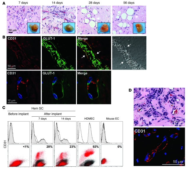
Figure 3. HemSCs form CD31+GLUT-1+ blood vessels in vivo.
(A) Clonal HemSCs were suspended in Matrigel and injected s.c. into nude mice. H&E sections of explants at 7, 14, 28, and 56 days with insets showing explants at the corresponding time points. Scale bar: 50 μm. Passage 9 clonal HemSCs were used. (B) Immunofluorescent staining of day 7 explants. Human CD31 (red) is shown on the left, followed by GLUT-1 (green), a merged image, and a phase contrast image. In the merged image, arrows point to cells along the lumen of the blood vessel that are double-labeled for human CD31 and GLUT-1. rbc in the lumen are also positive for GLUT-1 (green). In the phase image, arrows point to endothelial nuclei. The bottom row shows higher magnification images. Scale bars: 50 μm (top row); 10 μm (bottom row). (C) Flow cytometry of cells reisolated from Matrigel explants after 7 and 14 days in vivo. Cells were labeled with anti-human CD31 without permeabilization to detect surface-localized CD31. HemSCs before implantation were negative for CD31 (left panels). HDMECs and mouse ECs are shown as positive and negative control cells. (D) Retrieved CD31+ cells formed blood vessels in secondary recipient mice. Matrigel implants were removed after 14 days, sectioned, stained with H&E (top panel), and immunostained with anti-human CD31 (bottom panel). Vessel density was 78 ± 35 rbc-filled lumens/mm2. Scale bars: 50 μM.

