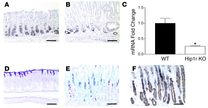
Figure 8. Loss of chief cells and expansion of an aberrant mucous neck cell population in Hip1r-deficient stomach.
(A and B) Chief cells in 2-month-old WT (A) and Hip1r-deficient (B) mice were identified by immunostaining paraffin sections with a polyclonal antibody to intrinsic factor. (C) Intrinsic factor mRNA abundance was determined by qRT-PCR analysis of RNA samples isolated from the corpus of 6-month-old WT and Hip1r-deficient mice. Data (mean ± SEM) were normalized to Gapdh expression in the same samples and reported as fold change relative to WT. n = 3. *P = 0.01 versus WT. (D–F) Expansion of aberrant mucous neck cells in Hip1r-deficient mice. Paraffin sections from 2-month-old WT (D) and Hip1r-deficient (E and F) mice were stained with PAS/Alcian blue (D and E) for neutral (pink) or acidic (blue) mucus or with GSII (F) to identify mucous neck cells. Scale bars: 100 μm.

