Figure 8.
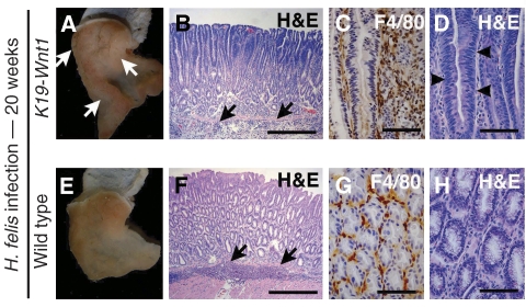
Tumour development in H. felis-infected K19-Wnt1 mouse stomach. K19-Wnt1 (A–D) and wild-type (E–H) mouse stomach infected with H. felis for 20 weeks. (A, E) Macroscopic photographs. The arrows in (A) indicate gastric tumours that developed in the infected K19-Wnt1 mice. H&E staining at low magnification (B, F) and high magnification (D, H). Immunostaining results for macrophage marker F4/80 (C, G). The arrows in (B, F) indicate submucosal infiltration. The arrowheads in (D) indicate dysplastic epithelial cells. Bars in (B, F) and (C, D, G, H) indicate 200 and 40 μm, respectively.

