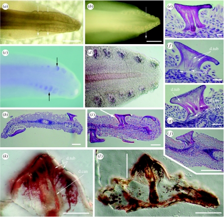
Figure 2.
Scale rows at S. canicula tail tip showing arrangement and histology compared to Ordovician scales from the Harding Sandstone. (a–d) Tail tip in left lateral view. (a) Transmitted light showing iterative scale pockets at stage 29 with an rostrocaudal stagger 9/10 dorsal and 8/9 ventral. (b) Incident light reflected from erupted scale surfaces showing tail shape (stage 33). (c) Photomacrograph (stage 32) and (d) Nomarski optics (DIC; stage 33) of whole mount in situ hybridization for Scshh showing expression patterns as iterative focal loci restricted to scale positions. (e–j) Scale histology in H&E stained vertical sections through the four scale rows (stage 33, DIC). (h) Scales either side of neural tube and notochord with large flat crowns and slender necks, (i) similar but ventral tail end (arrow, scale in (e)). (e) Mature scale showing two dentine canals and odontoblast cells below extended neck. (f) Young scale with shorter neck and odontoblast cell bodies in the dentine below the tubules. (g) More rostral, younger scale in (j) from dorsal row next to neural tube, with open base, several wide canals and odontoblast cell bodies below these. (k,l) Mineralized ground sections (DIC) of Ordovician shark microremains from Colorado (see also Sansom et al. 1996, fig. 1). (k) Higher magnification of crown dentine (line in (l)) of whole scale, mineral deposits in the spaces show tubular dentine with multiple branching from a wide central canal extending down through the extensive scale base. Abbreviations, d.can, dentine canal; d.tub, dentine tubule; od, odontoblasts. Scale bar, (a,c) 20 mm, (b) 500 μm, (d) 100 μm, (e–h) 10 μm and (i–l) 50 μm.

