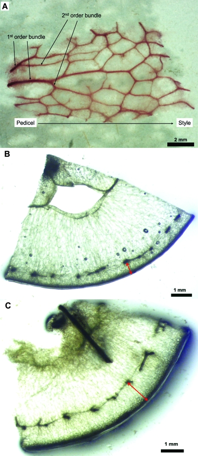
Fig. 1.
(A) Post-veraison peripheral vasculature network showing first-order vascular bundles (before any branching) and second-order vascular bundles. Basic fuchsin was infused using the wicking method, and the stained peripheral vasculature was dissected from the surrounding berry tissues. (B, C) Cross-sections near the berry equator at pre-veraison (DAA 48; B) and post-veraison (DAA 97; C) showing the distance between the epidermis and the peripheral vasculature (arrows).

