
Fig. 1.
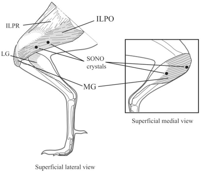
Schematic diagram of a guinea fowl hindlimb showing the anatomy of the iliotibialis lateralis pars postacetabularis (ILPO) and the medial head of the gastrocnemius (MG), highlighted in gray. Sonomicrometry (SONO) crystals are indicated by black circles. Crystals were implanted approximately midbelly in both muscles. EMG electrodes (not shown) were implanted adjacent to each pair of SONO crystals. The lateral gastrocnemius (LG) and iliotibialis lateralis pars preacetabularis (ILPR) are indicated for reference.