
FIG. 3. Diminished cortical F-actin and insulin-stimulated GLUT4 translocation in cells treated with chronic insulin are corrected by PIP2 replenishment.
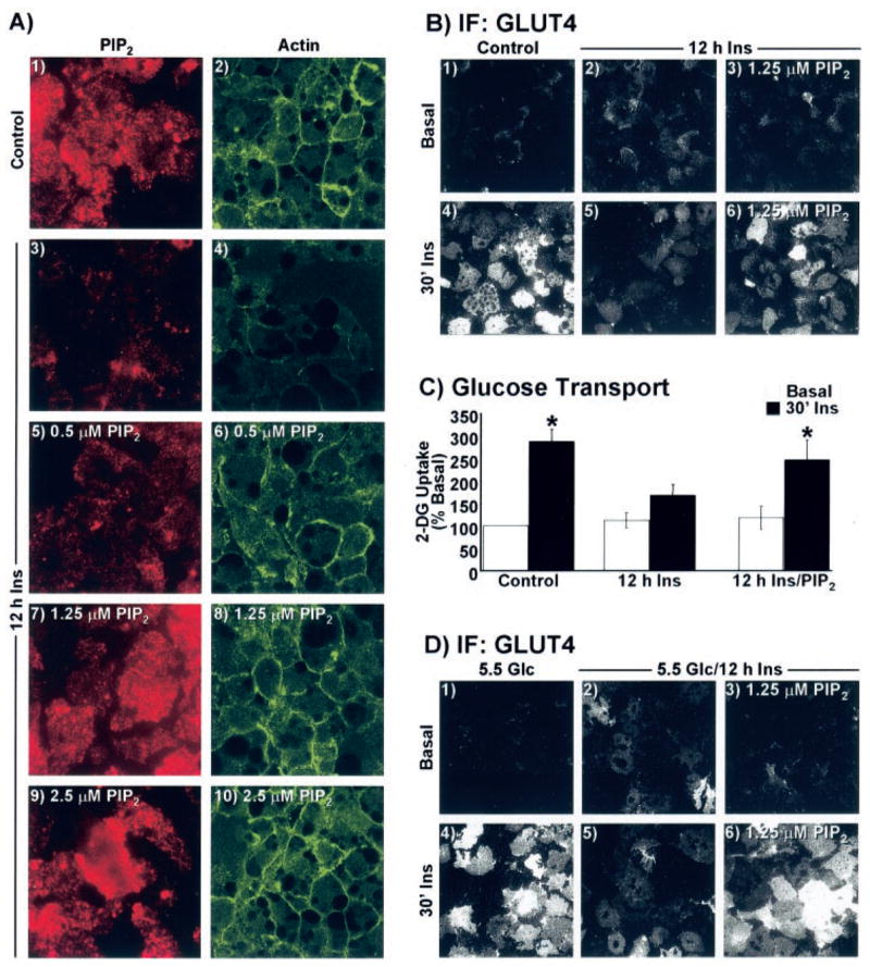
A–C, cells were treated overnight in the absence (Control) or presence of 5 nM insulin for 12 h. A, during the last 60 min of the 12-h period, the medium was replaced with the same medium enriched with either histone H1 (panels 1–4) or indicated concentrations of PIP2/histone H1 (panels 5–10). Plasma membrane sheets (panels 1, 3, 5, 7, and 9) or whole cells (panels 2, 4, 6, 8, and 10) were labeled for PIP2 (red) and actin (green), respectively. B, histone H1 (panels 1, 2, 4, and 5) or 1.25 μM PIP2 (panels 3 and 6) add-back incubations were performed as described for A, sheets were prepared, and GLUT4 immunofluorescence was assessed. C, 2-[3H]Deoxyglucose (2-DG) uptake was determined as described under “Experimental Procedures.” D, cells cultured in 5.5 mM glucose were treated overnight in the absence (5.5 Glc) or presence of 5 nM insulin (5.5 Glc/12 h Ins) for 12 h. Add-back treatment parameters and GLUT4 detection was performed exactly as described for A and B. All microscopic and camera settings were identical between groups and representative images from three independent experiments are shown.