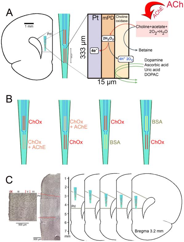
Figure 1.

(A) Schematic illustration depicting the main features, measurement scheme and dimensions of choline-sensitive microelectrode recording sites. Choline is generated by hydrolysis of newly released acetylcholine (ACh) by acetylcholinesterase (AChE). Choline is oxidized by choline oxidase (ChOx) immobilized onto the recording site. The resulting peroxide is detected amperometrically on Pt sites. m-PD electropolymerization serves to repel electroactive interferents including DA, AA, uric acid and DOPAC from the surface of the platinum recording sites. The left insert illustrates the placement and the relative dimensions of the four recording sites when placed into the medial prefrontal cortex (mPFC). (B) Schematic illustration of the enzyme coating combinations used in the present experiments. Recording sites were coated either with AChE+ChOx or with ChOx only. The position of the coating type on the electrode (upper versus lower pair) was systematically varied. For control experiments, recording sites on individual electrodes were coated with ChOx and BSA, respectively. (C) On the left, a coronal section stained for the visualization of AChE-positive fibers is shown. The prelimbic cortex (Prl) is indicated and shown at higher magnification (scales are 500 μm). The greater density of AChE-positive fibers in layer III is apparent. As shown on the schematic drawings on the right, KCl-evoked cholinergic signals were measured in layer III of the Prl in five places, spanning the dorsal-ventral extension of this region.
