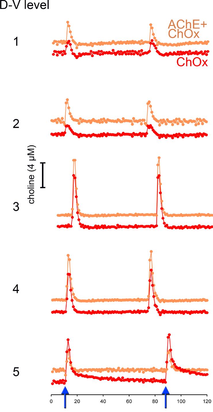
Figure 3.

Choline signals evoked by pressure-ejection of KCl (70 mM, 200 nL) via a micropipette whose tip was placed within 70-100 μm of the four recording sites. The orange trace shows choline signals recorded via AChE+ChOx-coated sites, while the red trace depicts responses via ChOx only sites. The effects of two successive pressure ejections of KCl are depicted (see arrows on abscissa on the bottom of the figure). Electrode/pipette assemblies were lowered by 200 μm after each series of KCl pressure ejections, for a total of 5 measures spanning the dorsal-ventral (D-V) extension of the prelimbic region. The response to KCl did not differ between the two types of recording sites or between placements (see Results for statistical findings).
