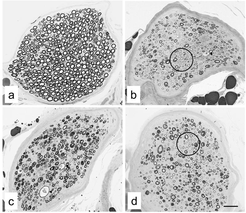
Fig. 2.
Examples of peroneal nerve biopsies from non-diabetic (a) and diabetic (b-d) cats from which endoneurial blood vessels were sampled. Note myelinated fiber density of peroneal nerve fascicles in non-diabetic cat (a) and the obvious myelinated fiber loss evident in diabetic cats (b-d). In diabetic cats, evidence of active demyelination was indicated by fibers with split and ballooned myelin sheaths (arrowheads in b and c), while remyelination was suggested by fibers with disproportionately thin myelin sheaths (circles in b and d) relative to those of fibers with comparable axonal diameters. Bar 73 μm (a), 46 μm (b), 55 μm (c) and 58 μm (d).

