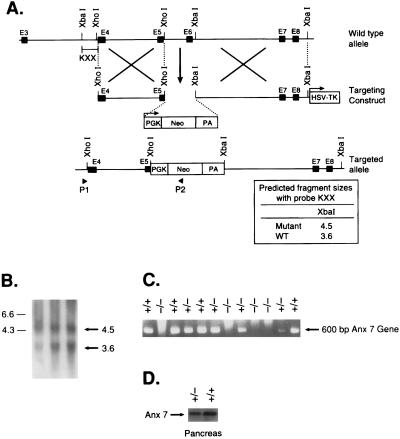
Figure 1.
Gene targeting strategy. (A) Restriction map of the mouse anx7 gene, the targeting vector, and the predicted structure of the locus after recombination. The top line represents the structure and partial restriction map from exon 3 to 9 of the wild-type allele of anx7. Exons are shown as black boxes. The middle line is the targeting plasmid in which a 900-bp XhoI and XbaI fragment is replaced with a 1.8-kb phosphoglycerate kinase (PGK)-neo-PA cassette and linearized at the unique NotI site. The dashed lines show the relationship of the two anx7 fragments in KSBX-pPNT to the genomic locus. The boxes represent the neo and HSV-tk genes, as labeled. The horizontal arrows show the phosphoglycerate kinase-1 promoters and their direction of transcription. The lowest line is the predicted structure of the targeted allele. The line marked KXX represents the 500-bp XhoI–XbaI fragment used for detection of homologous recombinants. The expected sizes of XbaI fragments that will hybridize to this probe is shown at lower right. (B) Southern blot analysis. Genomic DNA was digested with XbaI, blotted, and hybridized with KXX probe. The 3.6-kb fragment is derived from the wild-type allele, and the 4.5-kb fragment seen in lanes 1–3, representing 10–30 μg of genomic DNA from ES cells, is diagnostic of homologous recombination. (C) PCR analysis of genomic DNA from yolk sac of anx7(+/+), anx7(+/−), and anx7(−/−) embryos. The DNA (2 μl) was subjected to PCR analysis by using antisense oligonucleotide from the inserted neo cassette and sense oligonucleotide from genomic sequence outside the 2.0-kb 5′-flanking sequence in the targeting vector. (D) Immunoblot of anx7 extracted from pancreas of anx7(+/−) and control (+/+) mice. Pancreas from anx7(+/−) mice was harvested, frozen on dry ice, and then homogenized in boiling SDS buffer. Aliquots containing identical amounts of protein were separated by SDS/PAGE and transblotted to nitrocellulose, and ANX7 was visualized by Western blot analysis by using rabbit anti-human ANX7 primary antibody.

