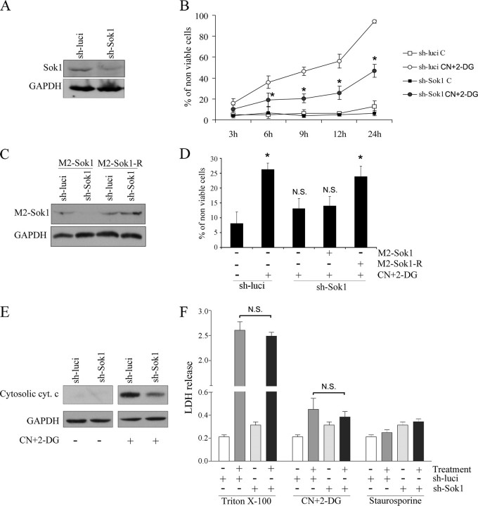
FIGURE 2.
Down-regulation of SOK1 protects cells from apoptosis induced by chemical anoxia. A, down-regulation of SOK1 by a hairpin RNA. HEK293 cells were transfected with a pSUPER-derived plasmid encoding a small hairpin RNA directed against SOK1 (sh-SOK1) or a control shRNA directed against firefly luciferase (sh-luci). 5 days later extracts were prepared, and a Western blot was performed to evaluate SOK1 down-regulation. GAPDH Western blot is shown as a loading control. B, inhibition of endogenous SOK1 delays cell death induced by chemical anoxia. HEK293 cells transfected with sh-SOK1 or sh-luci were either left untreated (C) or treated with sodium cyanide and 2-deoxyglucose (CN+2-DG) for the times indicated, and the percentage of non-viable cells was determined by the trypan blue exclusion assay. Shown is the mean ± S.D. of four independent experiments. *, p < 0.05 versus treated cells transfected with control shRNA. C, the SOK1R mutant is refractory to SOK1 shRNA. Cells transfected with sh-SOK1 or sh-luci were transfected again with either SOK1 wild-type or the shRNA-resistant SOK1R mutant. A Western blot was performed to assess the effect of sh-SOK1 on the different mutants of SOK1. D, the effects of sh-SOK1 are mediated by down-regulation of SOK1. Cells were then subjected to chemical anoxia for 6 h, and the percentage of dead cells assessed by trypan blue exclusion. Shown is the mean and S.D. of four independent experiments. p > 0.05 (N.S.) and p < 0.05 (*) versus untreated cells. E, SOK1 knockdown protects from the apoptotic cell death induced by chemical anoxia. HEK293 cells transfected with sh-SOK1 or sh-luci were either left untreated or treated with sodium cyanide and 2-deoxyglucose for 6 h. Cytochrome c (cyt. c) release was determined by Western blotting. GAPDH is shown as a loading control. F, SOK1 knockdown does not protect from the necrotic cell death induced by chemical anoxia. HEK293 cells transfected with sh-SOK1 or sh-luci were either left untreated or treated with sodium cyanide and 2-deoxyglucose for 6 h, and lactate dehydrogenase levels in the medium were measured. Triton X-100 was used as positive control, and staurosporine was used as a negative control of necrotic cell death. N.S., non-significant differences (p > 0.05) of treated cells with SOK1 shRNA versus treated cells with control shRNA.

