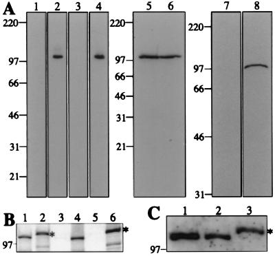
Figure 1.
Characterization of hCDC5 antisera. (A) Immunoblots of whole-cell protein extracts from NIH 3T3 (lanes 1, 2, and 5) and HeLa (lanes 3, 4, and 6–8) cells probed with preimmune (lanes 1 and 3), immune (lanes 2 and 4), and affinity-purified (lanes 5 and 6) antisera raised against the C terminus of hCDC5 (hCDC5C), and preimmune IgG (lane 7) and affinity-purified antisera (lane 8) raised against the N terminus of hCDC5 (hCDC5N). (B and C) Analysis of hCDC5 produced in vitro. hCDC5 or 6XHis/Myc-tagged hCDC5 proteins were produced in vitro in the presence (B) or absence (C) of TRAN35S-LABEL. In B, total products of the reactions (lanes 1 and 2), preimmune (lane 3) or anti-hCDC5C (lane 4) immunoprecipitates, and immunoprecipitates of the 6XHis/Myc-tagged hCDC5 with no primary antibody (lane 5) or 9E10 antibody (lane 6) were resolved by SDS/PAGE. Proteins were detected by fluorography. In C, an NIH 3T3 cell lysate (lane 1), hCDC5 produced in vitro (lane 2), and 6XHis/Myc-tagged hCDC5 produced in vitro (lane 3) were resolved by SDS/PAGE and immunoblotted with anti-hCDC5C serum. In both B and C, asterisks denote the 6XHis/Myc-tagged species of hCDC5. For all panels, numbers to the left indicate molecular mass (in kDa).

