Figure 3.
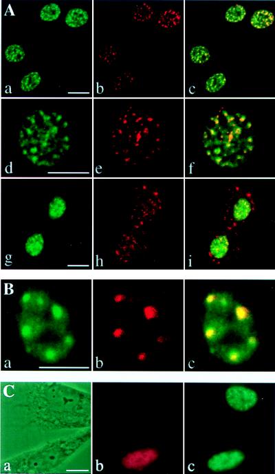
CDC5 is a component of the nuclear speckles. (A) NIH 3T3 cells were costained with affinity-purified hCDC5C antiserum and a monoclonal antibody against SC35. Fluorescein and Texas Red-conjugated secondary antibodies were used to detect the distribution of CDC5 (a, d, and g) and SC35 (b, e, and h), respectively. Confocal images of cells in interphase (a–f) and telophase (g–i) were obtained and merged (c, f, and i). Regions of colocalization are yellow in merged images. (B) Asynchronously growing tsBN2 cells were shifted to the nonpermissive temperature and costained with affinity-purified hCDC5C antiserum (a) and a monoclonal antibody to SC35 (b). The confocal images were merged in c. (C) Myc-tagged Clk/Sty kinase was transfected into NIH 3T3 cells. Phase contrast images (a) of cells costained with anti-Myc epitope monoclonal antibodies (9E10; b) and affinity-purified hCDC5C antiserum (c) at 24 h after transfection. (Bars = 10 μm.)

