
FIGURE 5.
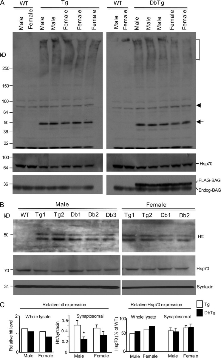
Expression of Hsp70 in male and female HD mice. A, immunoblots showing the expression of mutant htt, Hsp70, transgenic BAG1 (FLAG-BAG), and endogenous BAG (Endog-BAG) in the soluble lysates from the forebrain tissue of wild type (WT), N171-82Q (Tg), and double transgenic (DbTg) mice. The bracket indicates the stacking gel. The arrow indicates soluble transgenic htt, and the arrowhead indicates a nonspecific product that served as a loading control. The samples were probed with antibodies to htt (rabbit EM48, upper blot), Hsp70, and BAG1. Two to three male and female HD mice of each genotype were examined. B, Western blotting of synaptosomal fractions from 3- to 4-month-old male and female N171-82Q (Tg) or double transgenic (Db) mice expressing N171-82Q and BAG1. The blots were probed with mEM48 for transgenic htt (upper panel) and antibodies to Hsp70 (middle panel) and the synaptic protein synatxin (lower panel). Note that a smaller band or degraded product of mutant htt is more pronounced in synaptosomal fractions. C, densitometry analysis of the ratio of transgenic htt to the loading control in whole cell lysates or syntaxin in the synaptosomal fraction in N171-82Q (Tg) or double transgenic (DbTg) mice. The intact form or upper band of transgenic htt was used for quantification. The relative level of Hsp70 represents the percentage of Hsp70 level relative to the Hsp70 level in the wild-type mouse sample. The data (mean ± S.E.) were obtained from five to six samples of each group of two Western blotting experiments. *, p < 0.05.