
FIGURE 1.
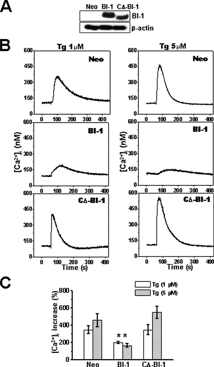
Internal stores of thapsigargin-releasable Ca2+ are reduced in BI-1 stable transfectants. A, expression of BI-1-HA tagged protein in HT1080-Neo, -BI-1, and -CΔ-BI-1 cells was compared by immunoblotting using an anti-HA antibody (upper panel). Lysates were normalized for total protein content (20 μg/lane). Blots were reprobed with anti-β-actin antibody to confirm equivalent loading (lower panel). B, HT1080-Neo, -BI-1, and -CΔ-BI-1 cells were loaded with Fura-2/AM, cultured in Ca2+-free medium, and incubated with 1 μm (left) or 5 μm (right) thapsigargin (Tg) at 1 min. Individual cells were imaged (n = 16), and average fluorescence intensity was recorded over time. C, the peak of Ca2+ release obtained in B was quantified and expressed as a percentage of the increase above base line prior to thapsigargin treatment (mean ± S.E. of four independent experiments). *, p < 0.05 by unpaired t test versus the percentage of increased cytoplasmic Ca2+ in the indicated concentration of thapsigargin in Neo cells.