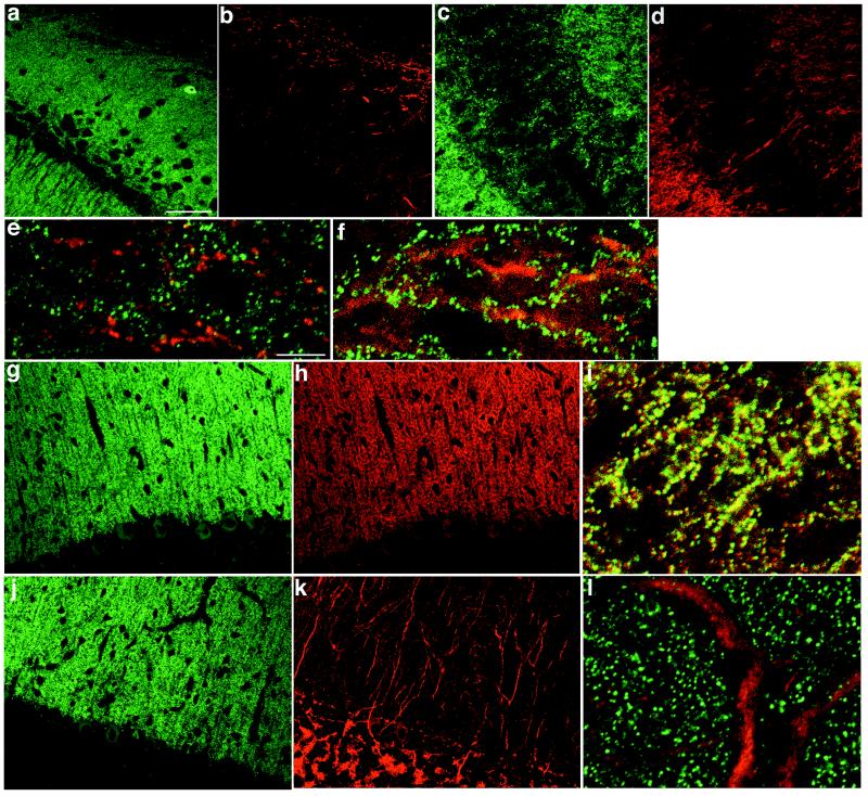
Figure 1.
Distribution of PSD-Zip45 and group 1 mGluRs in the rat brain. Sagittal sections of CA1 (a, b, and e) and CA3 (c, d, and f) regions of the hippocampus and of the cerebellum (g–l) were double labeled with a mixture containing mAb 126H (green; a, c, e–g, i, j, and l) and rabbit polyclonal antibodies for mGluR1 (red; b, e, h, and i) or mGluR5 (red; d, f, k, and l). e, f, i, and l are superimposed images. Scale bars = 50 μm (a–d, g, h, j, and k), 10 μm (e, f, i, and l).

