Figure 1.
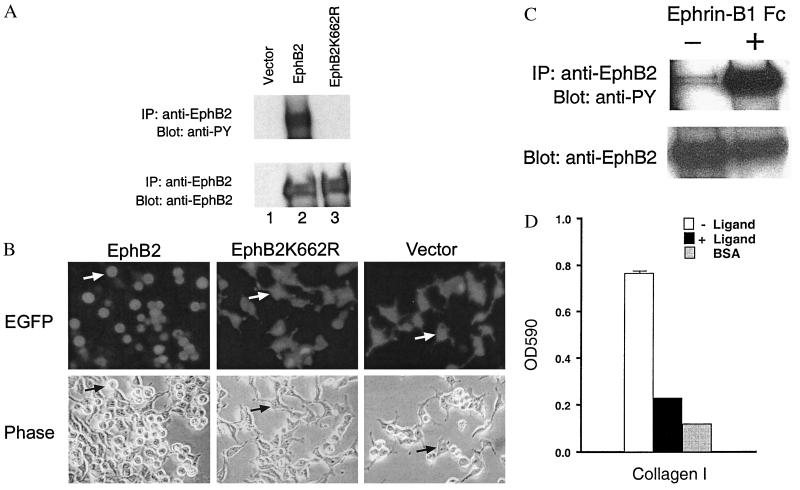
Activation of the EphB2 receptor and its effects on cell adhesion. (A) Activation of EphB2 in transiently transfected 293 cells. EphB2 was immunoprecipitated from cells transfected with EphB2, kinase-inactive EphB2K669, or pcDNA3. The immunoprecipitates (IP) were probed by immunoblotting as indicated. (B) Morphological changes caused by expression of activated EphB2 in 293T cells. An enhanced green fluorescent protein (EGFP) vector was cotransfected to identify the transfected cells. The transfected cells were shown to be alive by trypan blue exclusion. EphB2K662R and pcDNA3 vector were used as controls. (Upper) Fluorescence microscopy. (Lower) Phase-contrast microscopy of same fields. (×200.) (C) Activation of EphB2 by ephrin-B1 ligand. EphB2 was immunoprecipitated from NIH 3T3 cells that had been stably transfected with EphB2 and treated with soluble ephrin-B1 Fc ligand or left untreated. The immunoprecipitates were probed by immunoblotting as indicated. (D) Ligand stimulation of EphB2 decreases adhesion to collagen I in NIH 3T3 cells stably transfected with EphB2.

