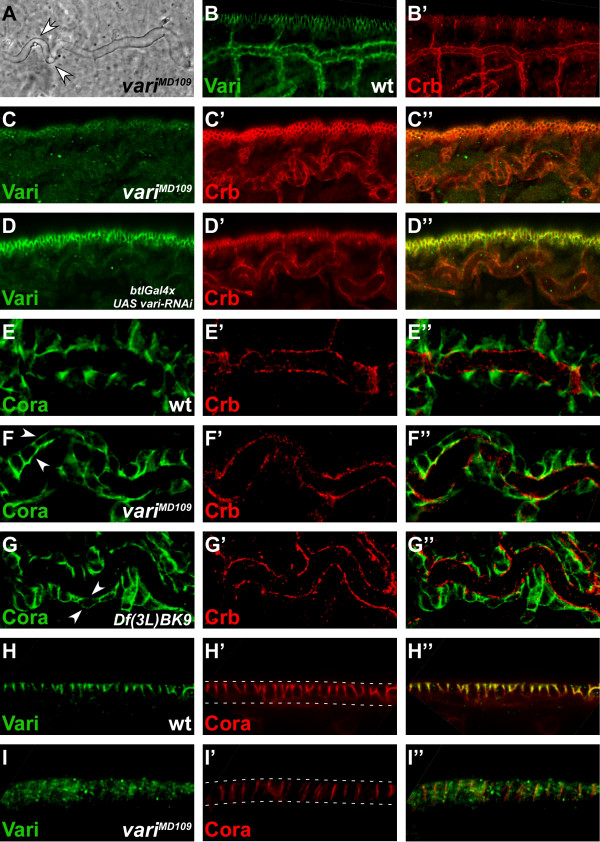
Figure 3.
Varicose is required for correct tracheal tube and epidermis formation. (A) Cuticle preparations of variMD109 mutant embryos exhibit convoluted tracheae (white arrows). (B, B') Wild-type embryo of stage 15 stained with anti-Vari (green) and anti-Crb (red). Vari localises at the SJ, basal of Crb. Wild-type tracheae appear straight in contrast to the convoluted tracheae in A. (C) variMD109 mutant embryo of stage 15 stained with anti-Vari (green) and anti-Crb (red). Vari is lost from the tracheae and the epidermis, while apical Crb is not affected. Tracheae appear convoluted. (D) Stage 15 embryo with targeted knockdown of vari in the tracheae of embryos by using btlGal4 (btlGal4>UAS vari-RNAi), stained with anti-Vari (green) and anti-Crb (red). Vari is reduced to background levels in the tracheae, but not affected in the epidermis. Apical localisation of Crb is not affected in the tracheae. (E) Dorsal tracheal trunk of a wild-type embryo of stage 15, stained with anti-Coracle (Cora; green) and anti-Crb (red). Cora localises in the SJ, basal to Crb. (F) Dorsal tracheal trunk of a variMD109 mutant embryo of stage 15, stained with anti-Cora (green) and Crb (red). Cora is delocalised to apical and basal sites (white arrows), whereas Crb remains in its apical position. (G) Dorsal tracheal trunk of a Df(3L)BK9 mutant embryo of stage 15, in which the NrxIV locus is deleted, stained with anti-Cora (green) and anti-Crb (red). As in variMD109 mutant embryos, Cora becomes mislocalised to apical and basal positions (white arrows) in the absence of NrxIV, while apical localisation of Crb is not affected. (H) Epidermis of a wild-type embryo of stage 15, stained with anti-Vari (green) and anti-Cora (red). Both proteins are co-localised at the SJ. (I) variMD109 mutant embryo of stage 15, stained with anti-Vari (green) and anti-Cora (red). The amount of Cora is reduced and the remaining Cora protein is mislocalised along the whole lateral membrane. In B-D and H-I apical is up. White dotted lines in H' and I' mark the apical and basal side of the epithelial cells, respectively.

