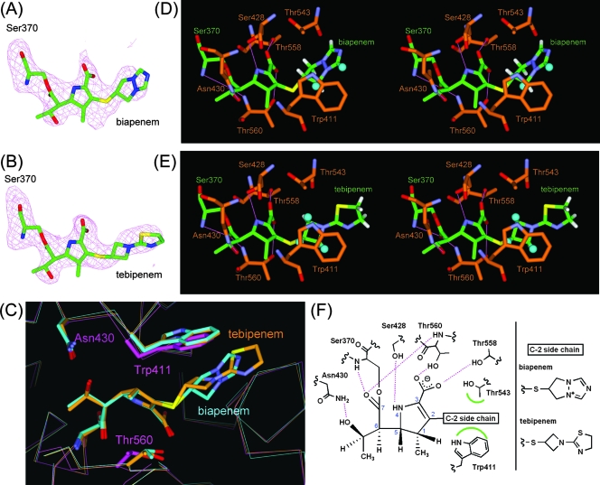
FIG. 3.
PBP1A-carbapenem complex structures. (A and B) Fo − Fc omit electron density maps calculated for the structures omitting the carbapenem moiety covalently attached to Ser370 in molecule 1 of PBP 1A in complex with biapenem and tebipenem. The maps and models are displayed in the same manner as for Fig. 2A and B. (C) Superpositioned active-site regions of the uncomplexed (magenta) and biapenem (light blue)- and tebipenem (orange)-complexed structures. The carbapenems and side chains of selected residues are shown as thick sticks. Atoms are colored as described for Fig. 2C. (D and E) Stereo view of the active sites of PBP 1A in complex with biapenem and tebipenem, respectively. Atoms are colored as described for Fig. 2A and B, except that the carbon atoms of PBP 1A are orange. Hydrogen atoms of the C-2 side chain and hydrogen bonds are displayed in the same manner as for Fig. 2D and E. (F) Schematic diagram of the carbapenem binding mode. Hydrogen bonds are represented by broken lines (magenta), and hydrophobic contacts are shown as arcs (green). The atomic numbering scheme of the carbapenem skeleton is also shown.

