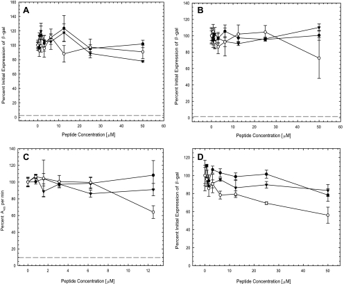
FIG. 4.
Antiviral properties of β-peptides. To determine the step in infection blocked by the β-peptides, the time of introduction of β-peptides in four assays was varied as described in Materials and Methods. (A) Inactivation of virions in solution. Virus was incubated with various concentrations of the β-peptides at 37°C for 1 h. The β-peptide-virus mixtures were then diluted 1,000-fold, and the titer of infectious virus was determined. The bKLA peptide (30 μM) was included as a positive control for virus inactivation in solution (dashed line). Infectivity was determined after 6 h at 37°C by measuring β-galactosidase (β-gal) activity. (B) Induction of cellular resistance to infection. Vero cells were incubated with various concentrations of the β-peptides at 37°C for 1 h. β-Peptides were removed, and the cells were rinsed and infected with virus for 1 h at 37°C. The TAT-Cd peptide (100 μM) was included as a positive control for induction of resistance to infection (dashed line). Infectivity was determined after 6 h at 37°C by measuring β-galactosidase activity. (C) Viral attachment. Vero cells were cooled at 4°C for 30 min. The cultures were then incubated with virus in the presence of various concentrations of the β-peptides at 4°C for 1 h. The cultures were fixed with 4% paraformaldehyde and blocked with 3% BSA in medium for 1 h at room temperature. Primary and secondary antibodies were polyclonal rabbit anti-HSV-1 antibody (Dako North America, Inc.) and goat anti-rabbit IgG conjugated with alkaline phosphatase (Sigma-Aldrich), respectively. The kinetics of absorbance at 405 nm was recorded as a measure of viral attachment. Heparin (5 μM) was included as a positive control for inhibition of viral attachment (dashed line). (D) Postentry inhibition. Vero cells were infected with virus for 1 h at 37°C. The cultures were then rinsed, treated with low-pH citrate buffer (pH 3.0) to inactivate extracellular virus, rinsed again, and refed with medium containing various concentrations of the β-peptides. Infectivity was determined after 6 h at 37°C by measuring β-galactosidase activity. The negative control was a sample without β-peptide (set as 100%) in all assays. Each data point and error bar represent the mean ± standard deviation of results for triplicate samples. ○, peptide 1; •, peptide 2; ▾, peptide 3.

