Figure 1.
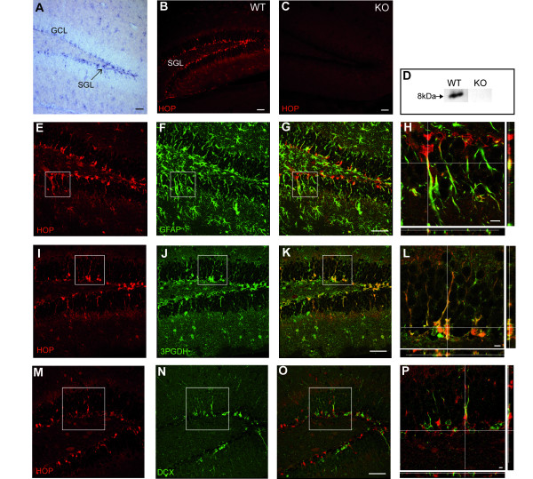
HOP is expressed by SGL radial astrocytes. (a) In situ hybridization shows HOP mRNA expression in numerous DG cells with a prominent labeling in the SGL (arrow), the neurogenic region for the production of granule cells destined to the granule cell layer (GCL). (b) Immunohistochemical detection of HOP protein in the DG, especially in the SGL. WT, wild type. (c) HOP immunohistochemical staining is absent in knock out (KO) mice, demonstrating HOP antibody specificity. (d) Western blot shows an 8 kDa protein from hippocampal wild-type extracts, which is absent in the knock out extracts. (e-h) Double-staining with HOP and GFAP antibodies, illustrating the co-expression of the two proteins. (h) Higher magnification of the boxed area in (e-g) with orthogonal projections along the z confocal stack at the x and y levels indicated by the intersecting white lines. (i-l) Double-staining with HOP and 3PGDH antibodies, showing that HOP immunoreactive cells are 3PGDH+. (l) Higher magnification of the boxed area with orthogonal projections. (m-p) Double-staining with HOP and DCX, showing that they are expressed by different SGL cells. (p) Higher magnification of the boxed area with orthogonal projections. The y projection shows, in particular, that DCX+ processes are closely juxtaposed to HOP+ processes but are distinct. Scale bars: (a-f, e-g, i-k, m-p), 50 μm; (h, l, p), 10 μm.

